A collaborative work between CMRR and UMRAM resulted in a manuscript entitled “In vivo human head MRI at 10.5T: A radiofrequency safety study and preliminary imaging results.” In this study, the first human brain images obtained from a 10.5T system are shown. 10.5T is currently the highest field strength full-body imaging system in the world and housed at CMRR in the University of Minnesota. The first author of the paper is Alireza Sadeghi‐Tarakameh who is a PhD student of Ergin Atalar. The senior author of the article, Yiğitcan Eryaman is an assistant professor at CMRR and received his PhD degree from Bilkent University. The other authors of the paper are Lance DelaBarre, Russell L. Lagore, Angel Torrado-Carvajal, Xiaoping Wu, Andrea Grant, Gregor Adriany, Gregory J. Metzger, Pierre-Francois Van de Moortele, and Kamil Ugurbil of CMRR.
Abstract
Purpose
The purpose of this study is to safely acquire the first human head images at 10.5 T.
Methods
In order to ensure safety of subjects, we validated the electromagnetic (EM) simulation model of our coil. We obtained quantitative agreement between simulated and experimental B1+ and specific absorption rate (SAR). Using the validated coil model, we calculated radiofrequency (RF) power levels to safely image human subjects. We conducted all experiments and imaging sessions in a controlled RF safety lab and the whole-body 10.5T scanner in the Center for Magnetic Resonance Research (CMRR).
Results
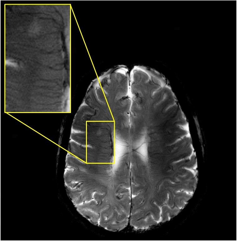
Quantitative agreement between the simulated and experimental results was obtained including S-parameters, B1+ maps, and SAR. We calculated peak 10 g average SAR using four different realistic human body models for a quadrature excitation and demonstrated that the peak 10 g SAR variation between subjects was less than 30%. We calculated safe power limits based on this set and used those limits to acquire T2- and T2*-weighted images of human subjects at 10.5 T.
Conclusion
In this study, we acquired the first in vivo human head images at 10.5 T using an eight-channel transmit/receive (Tx/Rx) coil. We implemented and expanded a previously proposed workflow to validate the EM simulation model of the eight-channel Tx/Rx coil. Using the validated coil model, we calculated RF power levels to safely image human subjects.