CMRR ve UMRAM arasındaki işbirliğine dayalı bir çalışma “10.5T’de in vivo insan kafası MRG: Bir radyofrekans güvenlik çalışması ve ön görüntüleme sonuçları” başlığı ile yayımlandı. Bu çalışmada, 10.5T sisteminden elde edilen ilk insan beyni görüntüleri gösterilmektedir. 10.5T şu anda dünyadaki en yüksek alan gücü olan tam vücut görüntüleme sistemidir ve Minnesota Üniversitesi’ndeki CMRR’de bulunmaktadır. Makalenin ilk yazarı Prof.Dr.Ergin Atalar‘ın doktora öğrencisi olan Alireza Sadeghi ‐ Tarakameh‘dir. Makalenin kıdemli yazarı Yiğitcan Eryaman, CMRR’de yardımcı doçent olarak görev yapmaktadır ve Bilkent Üniversitesi’nden doktora derecesini almıştır. Makalenin diğer yazarları ise Lance DelaBarre, Russell L. Lagore, Angel Torrado-Carvajal, Xiaoping Wu, Andrea Grant, Gregor Adriany, Gregory J. Metzger, Pierre-Francois Van de Moortele ve CMRR’den Kamil Uğurbil’dir.
Abstract
Purpose
The purpose of this study is to safely acquire the first human head images at 10.5 T.
Methods
In order to ensure safety of subjects, we validated the electromagnetic (EM) simulation model of our coil. We obtained quantitative agreement between simulated and experimental B1+ and specific absorption rate (SAR). Using the validated coil model, we calculated radiofrequency (RF) power levels to safely image human subjects. We conducted all experiments and imaging sessions in a controlled RF safety lab and the whole-body 10.5T scanner in the Center for Magnetic Resonance Research (CMRR).
Results
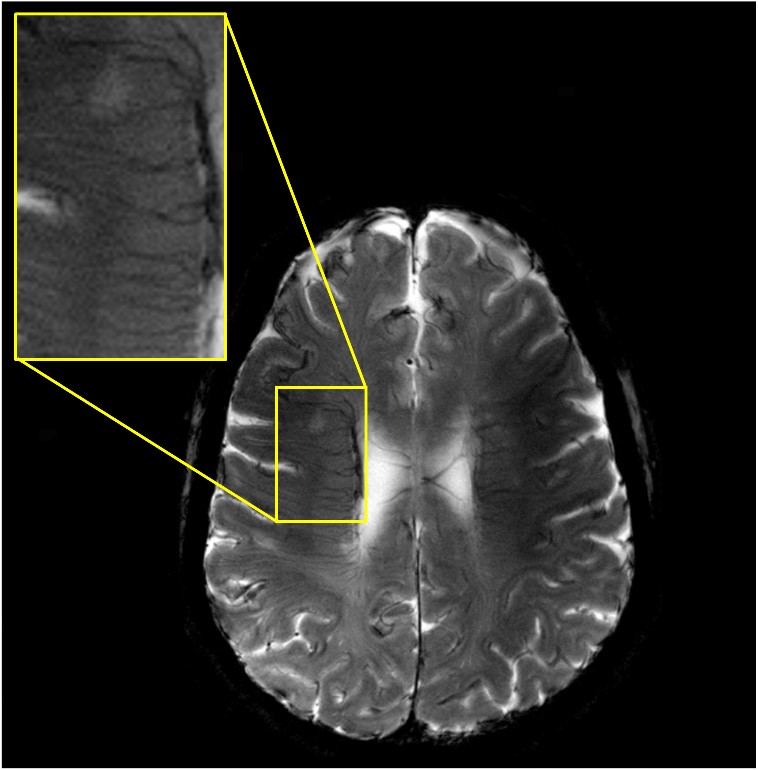
Quantitative agreement between the simulated and experimental results was obtained including S-parameters, B1+ maps, and SAR. We calculated peak 10 g average SAR using four different realistic human body models for a quadrature excitation and demonstrated that the peak 10 g SAR variation between subjects was less than 30%. We calculated safe power limits based on this set and used those limits to acquire T2- and T2*-weighted images of human subjects at 10.5 T.
Conclusion
In this study, we acquired the first in vivo human head images at 10.5 T using an eight-channel transmit/receive (Tx/Rx) coil. We implemented and expanded a previously proposed workflow to validate the EM simulation model of the eight-channel Tx/Rx coil. Using the validated coil model, we calculated RF power levels to safely image human subjects.