

UMRAM faculty member, Dr. Tolga Cukur has received the Mustafa Parlar Research Incentive Award for his work on biomedical imaging technology for radiological and neuroscientific applications. This award is given yearly by the Middle East Technical University Parlar Foundation to the highly selected young researchers. This year four Bilkent University faculty, Ali Jaili, Cem Tekin, Onur Tokel and Tolga Cukur received the award. The award ceremony will be held in METU Culture and Convention Center on December 19, 2019.
News
The journal articles by UMRAM researchers published in October 2026.
- Understanding Older Adults’ And Caretakers’ Attitudes Toward Social Robots Using Natural Language Processing and Thematic Analysis, International Journal of Social Robotics. Authors: Begüm Çonka, Burcu Aysen Urgen, Yasemin Afacan
- A recurrent neuronal model for the effect of predictions on sensory processes, Scientific Reports. Authors: Buse Merve Ürgen, Hilal İrem Baştürk, Hüseyin Boyacı

Open Positions/ Msc or PhD
Institute We offer a funded PhD. or Msc. scholarships on Medical Image Acquisition and Reconstruction at the National Magnetic Resonance Center. The position will be based in Bilkent University National Magnetic Resonance…
Institute
We offer a funded PhD. or Msc. scholarships on Medical Image Acquisition and Reconstruction at the National Magnetic Resonance Center. The position will be based in Bilkent University National Magnetic Resonance Research Center (UMRAM). The position holder will be in close collaboration with Istanbul Technical University.
Medical Image Reconstruction
Several recent advances have led to more efficient ways in which MRI can be reconstructed. The most prominent example of these is compressed sensing MRI, which allows the reconstruction of MRI from under-sampled k-space data. The reconstruction of the MRI requires solving a non-linear optimization problem. For medical image reconstruction, machine learning can play a key role. The project aims to exploit recent advances in machine learning, in particular in the area of representation learning to learn representations and priors directly from raw data that are optimal for recovery of images (or even for the reconstruction of clinically useful information). The key tasks for the project are:
- Acquisition of raw cardiac MR data
- Reconstruction of cardiac MRI from under-sampled k-space data.
- Development of deep learning based reconstruction algorithms leveraging temporal dependencies in cardiac MRI
Funding & Details
PhD position is fully funded (4500TL/month) by The Scientific and Technological Research Council of Turkey (TÜBİTAK). In addition, MSc. position might also be considered with funding. The prospective students will be able to attend to prestigious international conferences and spend some time in top international institutes. The candidates must be enrolled in a graduate program in Turkey and can start to work from February 2020. We encourage applications from underrepresented groups.
Required Skills
We are looking for highly motivated students with an academic background in Physics, Computer Science or Engineering. The following skills are required:
- Experience with Python and deep learning libraries (e.g. Pytorch, Tensorflow or Keras).
- Motivation in machine learning and computer vision problems/algorithms.
- Enthusiasm to work on clinical challenges.
- Good academic writing skills and English communication skills.
In vivo human head MRI at 10.5T: A radiofrequency safety study and preliminary imaging results
Our PhD student Alireza Sadeghi‐Tarakameh, Prof. Dr. Ergin Atalar and our PhD alumni Yiğitcan Eryaman published a paper titled “In vivo human head MRI at 10.5T: A radiofrequency safety study and preliminary imaging results” with Lance DelaBarre, Russell L. Lagore, Angel Torrado‐Carvajal, Xiaoping Wu, Andrea Grant, Gregor Adriany, Gregory J. Metzger, Pierre-Francois Van de Moortele, and Kamil Ugurbil in Magnetic Resonance in Medicine.
Abstract
PURPOSE:
The purpose of this study is to safely acquire the first human head images at 10.5T.
METHODS:
To ensure safety of subjects, we validated the electromagnetic simula-tion model of our coil. We obtained quantitative agreement between simulated and experimental B1+ and specific absorption rate (SAR). Using the validated coil model, we calculated radiofrequency power levels to safely image human subjects. We con-ducted all experiments and imaging sessions in a controlled radiofrequency safety lab and the whole-body 10.5T scanner in the Center for Magnetic Resonance Research
RESULTS:
Quantitative agreement between the simulated and experimental results was obtained including S-parameters, B+1 maps, and SAR. We calculated peak 10 g average SAR using 4 different realistic human body models for a quadrature excita-tion and demonstrated that the peak 10 g SAR variation between subjects was less than 30%. We calculated safe power limits based on this set and used those limits to acquire T2- and T∗2-weighted images of human subjects at 10.5T.
CONCLUSIONS:
In this study, we acquired the first in vivo human head images at 10.5T using an 8-channel transmit/receive coil. We implemented and expanded a previously proposed workflow to validate the electromagnetic simulation model of the 8-channel transmit/receive coil. Using the validated coil model, we calculated radiofrequency power levels to safely image human subjects.
MAGNETIC RESONANCE IN MEDICINE
DOI: 10.1002/mrm.28093
Wireless control of induced radiofrequency currents in active implantable medical devices during MRI

Our PhD alumni Dr. Volkan Açıkel, personnel alumni Berk Silemek and Prof. Dr. Ergin Atalar published a paper titled “Wireless control of induced radiofrequency currents in active implantable medical devices during MRI” in Magnetic Resonance in Medicine.
Abstract
Purpose: To introduce a prototype active implantable medical device (AIMD) for which the induced radiofrequency currents can be controlled wirelessly. Methods: The modified transmission line method is used to formulate how the lead-case impedance of an AIMD affects the temperature rise around the electrode. A prototype AIMD is designed with the aim of controlling the unwanted temperature rise around its electrode during an MRI examination by altering the impedance between the lead and the case of the implant. MRI experiments were conducted with this prototype implant, which also has a built-in temperature sensor at its electrode. During the experiment, the implant’s lead-case impedance was controlled using Bluetooth communication with a remote computer, and the lead tip temperature was recorded. Results: Ten different lead-case impedance values and their corresponding tip temperature rises were examined during MRI experiments. The experimental results confirmed that the tip temperature rise can be controlled by varying the lead-case impedance wirelessly. Conclusion: The feedback from the temperature at the AIMD tip, together with variable lead-case impedance, enables control of the safety profile of the AIMD during an MRI examination.
MAGNETIC RESONANCE IN MEDICINE
DOI: 10.1002/mrm.28089
UMRAM Will Be Evaluated Within the Scope of Law No. 6550 on Supporting Research Infrastructures
UMRAM (National Magnetic Resonance Research Center) will be evaluated to qualify as a legal entity within the scope of Law No. 6550 on Supporting Research Infrastructures.
TÜBİTAK officials and panelists are going to visit UMRAM on 24 December 2019 for evaluation. During the visit, UMRAM’s existing infrastructure will be evaluated and future plans will be discussed. Preparations for this visit are underway in UMRAM. In addition to scientific activities (publications, patents, etc.) after UMRAM launches, services for users will probably come to the fore most importantly.
If UMRAM is included in 6550, it is expected that there will be a significant increase in its scientific activities and services.
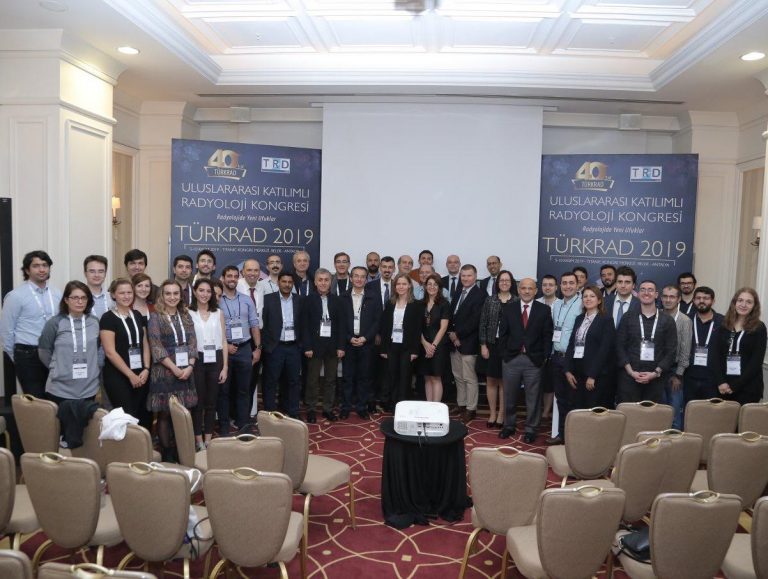
New Medical Imaging Techniques: From Laboratory to Clinics: A session in TURKRAD 2019
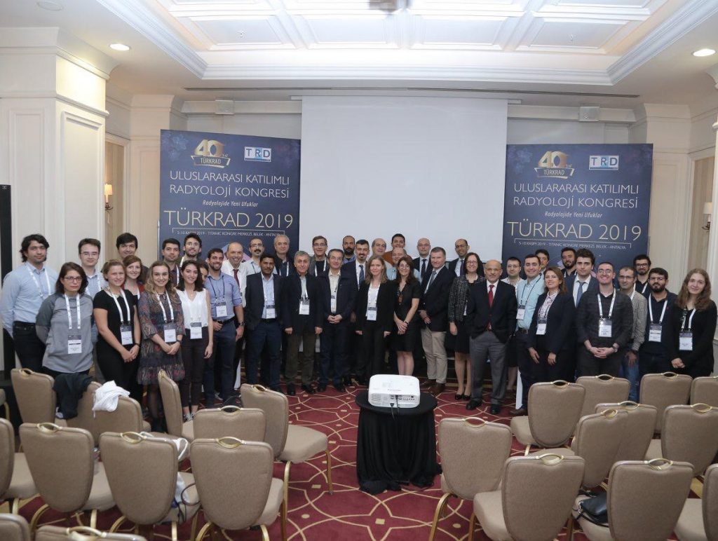
Annual Radiology Congress TURKRAD 2019 organized by Turkish Society of Radiology was held between 5th and 10th November 2019 at the Titanic Convention Center in Belek, Antalya. 40th National Radiology Congress was held with International Participation. There were important participants from Bilkent University, UMRAM and ASELSAN.
In the congress, a workshop was organized bringing together researchers working in imaging technologies with the support of ASELSAN. “New Medical Imaging Techniques: From Laboratory to Clinic” course was held on November 7 and 8, 2019.
On the first day of this course, Kader Karlı Oğuz, Ömer Demirkaya and Kamil Uğurbil made introductory speeches. Then the sessions of “Domestic Imaging Devices”, “Deep Learning”, “Innovative Imaging Techniques 1”, “MRI Data Processing”, “Innovative Imaging Techniques 2 were held.
Session: “Domestic Imaging Devices”
The moderators of the session were Ergin Atalar and Tuncay Hazırolan from UMRAM.
Speakers from UMRAM and ASELSAN were Ergin Atalar, Volkan Açıkel, Manouchehr Takrimi, Ehsan Kazemivalipour and Reza Babaloo.
Session: “Deep Learning”
Tolga Çukur was among the moderators of the session.
Speakers from UMRAM were Tolga Çukur, Abdullah Ömer Arol, Bilal Taşdelen and Süheyl Taraghinia.
Session: “Innovative Imaging Techniques 1″
The moderators of the session were Emine Ülkü Sarıtaş and Yusuf Ziya İder.
Speakers from Bilkent University and UMRAM were Yusuf Ziya İder, Murat Eyüboğlu, Emine Ülkü Sarıtaş and Ahmet Rahmetullah Çağıl.
Session: “MRI Data Processing”
Murat Eyüboğlu was among the moderators of the session.
Speakers from UMRAM were Tolga Çukur and Laleh Eskhandarian.
Session: “Innovative Imaging Techniques 2″
Speakers from UMRAM were Cemre Arıyürek, Ali Alper Özaslan and Ecrin Yağız.
You can reach the detailed program here.

TURKRAD 2019 PANEL “MADE IN TURKEY”: İBRAHİM BEKAR and ERGİN ATALAR
In the Annual Radiology Congress TURKRAD 2019 organized by Turkish Society of Radiology there was a debate on the production on medical imaging equipment in Turkey.
In the session, “MADE IN TURKEY”, which was held on Wednesday, November 6, 2019, ASELSAN Vice President Dr. İbrahim Bekar talked on “Radiology Industry in Turkey” and Bilkent University. UMRAM Director Prof. Dr. Ergin Atalar‘s talk was entitled “R&Ds in Radiology Technologies in Turkey”. Later there was a long discussion on the topic.
Currently, Turkey does not have high-tech medical industry. UMRAM is committed working with the industry in order to enable them to produce medical imaging equipment. Currently, a prototype MRI scanner is under development together with ASELSAN.

Interview with Dr. Kader Karli Oguz
Siemens made an interview with Dr. Oguz for their magazine’s November 2019 issue. You can reach the interview here.
Dr. Kader KARLI OGUZ, the Professor of Radiology at Hacettepe and faculty member of UMRAM, was the program chair of TURKRAD 2019, the 40th Radiology Congress.
Annual Radiology Congress TURKRAD 2019 organized by Turkish Society of Radiology was held between 5th and 10th November 2019 at the Titanic Convention Center in Belek, Antalya. 40th National Radiology Congress was held with International Participation.
Mersin University is looking for 5 PhD students for TUBITAK BIDEB 2232
Mersin University is looking for 5 PhD students for the project “Rescue Food Reward in Obesity with Neuro-modulation of gut-brain signals” funded under Tubitak BIDEB 2232 - Uluslararası Lider Araştırmacılar Programı. The…
Mersin University is looking for 5 PhD students for the project “Rescue Food Reward in Obesity with Neuro-modulation of gut-brain signals” funded under Tubitak BIDEB 2232 – Uluslararası Lider Araştırmacılar Programı. The duration of this project is 3 years. Starting date to be determined, preferably in the last quarter of 2019.
3 PhD students will enroll at the graduate program in Anatomy at Mersin University and 2 PhD students that are already in a graduate program elsewhere can perform studies at Bilkent University. The scholarship will be 4500 TL monthly. Equipment and funds for studies are present. Elective course work at both Bilkent and Mersin Universities is possible for all students.
More on the research of the project coordinator can be found here: https://www.mariaveldhuizen.com/
Please email the project coordinator at maria.veldhuizen@yale.edu for more information.
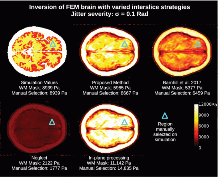
”Fast Robust Dejitter and Interslice Discontinuity Removal in MRI Phase Acquisitions: Application to Magnetic Resonance Elastography” by Cemre Ariyurek as co-author

Our PhD student Cemre Ariyurek has contributed to and published a paper as co-author, entitled “Fast Robust Dejitter and Interslice Discontinuity Removal in MRI Phase Acquisitions: Application to Magnetic Resonance Elastography”, with her collaborators Eric Barnhill, Mila Nikolova, Florian Dittmann, Jurgen Braun and Ingolf Sack.
The authors were honored to have co-authored
this publication with the great mathematician Mila Nikolova and dedicate it to
her memory.
Abstract:
MRI phase contrast imaging methods that
assemble slice-wise acquisitions into volumes can contain interslice phase
discontinuities (IPDs) over the course of the scan from sources, including
unavoidable physiological activity. In magnetic resonance elastography (MRE),
this can alter wavelength and tissue stiffness estimates, invalidating the
analysis. We first model this behavior as jitter along the z-axis of the phase
of 3D complex-valued wave volumes. A two-step image processing pipeline is then
proposed that removes IPDs. First, constant slicewise phase shift is removed
with a novel, non-convex dejittering algorithm. Then, regional physiological
noise artifacts are removed with novel filtering of 3D wavelet coefficients.
Calibration of two pipeline coefficients, the dejitter parameter α and the
wavelet band high-pass coefficient ωc , was first performed on a finite-element
method brain phantom. A comparative investigation was then performed, on a
cohort of 48 brain acquisitions, of four approaches to IPDs: 1) the proposed
method; 2) a “control” condition of neglect of IPDs; 3) an
anisotropic wavelet-based method; and 4) a method of in-plane (2D) processing.
The present method showed medians of [Formula: see text] Pa for a
multifrequency wave inversion centered at 40 Hz which was within 6% of methods
3) and 4), while neglect produced [Formula: see text] estimates a mean of 17%
lower. The proposed method reduced the value range of the cohort against
methods 3) and 4) by 29% and 31%, respectively. Such reduction in variance
enhances the ability of brain MRE to predict subtler physiological changes. Our
theoretical approach further enables more powerful applications of fundamental
findings in noise and denoising to MRE.
Barnhill, E., Nikolova, M., Ariyurek, C.,
Dittmann, F., Braun, J., & Sack, I. (2019). Fast Robust Dejitter and
Interslice Discontinuity Removal in MRI Phase Acquisitions: Application to
Magnetic Resonance Elastography. Ieee Transactions on Medical
Imaging, 38(7), 1578–1587. http://doi.org/10.1109/TMI.2019.2893369
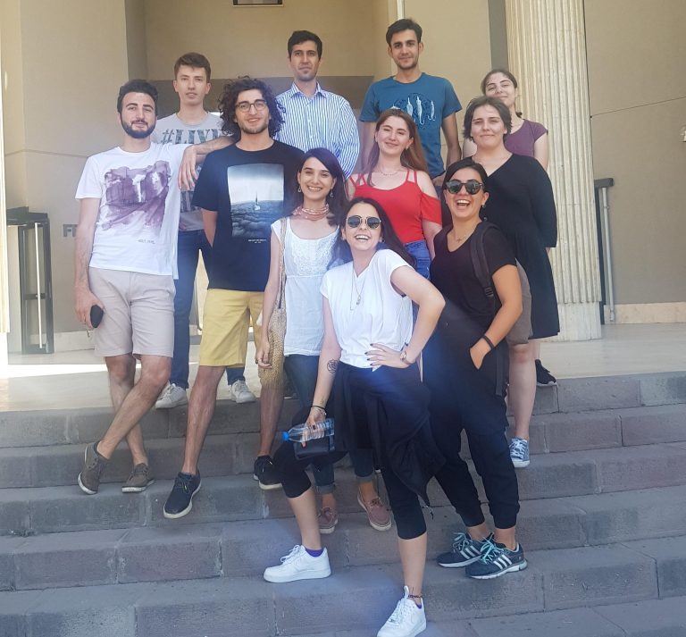
2018-2019 summer term interns of UMRAM graduated
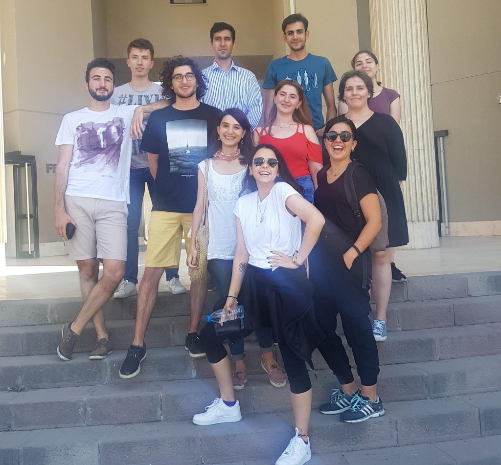
In addition to Bilkent University, Koc University, METU, Hacettepe University students from different undergraduate departments completed their summer internship at our center. After completing their compulsory internship, some of them wanted to continue in the studies voluntarily for a while.
By the August 1, we said goodbye to our interns. We thank them for their efforts and contributions and wish success for the rest of their lives.
Disentangling simultaneous changes of surface and illumination
Our faculty member Ass.Prof. Katja Doerschner has published a paper with Robert Ennis from Justus-Liebig-Universitaet Giessen. Abstract: Retinally incident light is an ambiguous product of spectral distributions of light in the environment…
Our faculty member Ass.Prof. Katja Doerschner has published a paper with Robert Ennis from Justus-Liebig-Universitaet Giessen.
Abstract:
Retinally incident light is an ambiguous product of spectral distributions of light in the environment and their interactions with reflecting, absorbing, and transmitting materials. An ideal color constant observer would unravel these confounded sources of information and account for changes in each factor. Scene statistics have been proposed as a way to compensate for changes in the illumination, but few theories consider changes of 3-dimensional surfaces. Here, we investigated the visual system’s capacity to deal with simultaneous changes in illumination and surfaces. Spheres were imaged with a hyperspectral camera in a white box and their colors, as well as that of the illumination were varied along “red-green” and “blue-yellow” axes. Both the original hyperspectral images and replica scenes rendered with Mitsuba were used as stimuli, including rendered scenes with Glavens (Acta Psychologica, 2009, 132, 259–266). Observers viewed sequential, random pairs of our images, with either the whole scene, only the object, or only a part of the background being present. They judged how much the illuminant and object color changed on a scale of 0–100%. Observers could extract simultaneous illumination and reflectance changes when provided with a view of the whole scene, but global scene statistics did not fully account for their behavior, while local scene statistics improved the situation. There was no effect of color axis, shape, or simulated vs. original hyperspectral images. Observers appear to be making use of various sources of local information to complete the task.
Ennis, R., & Doerschner, K. (2019). Disentangling simultaneous changes of surface and illumination. Vision Research, 158, 173–188. http://doi.org/10.1016/j.visres.2019.02.004